Monday, July 31, 2023

End-of-season assessment of sclerotinia stem rot of canola (PCDMN)

The spring and summer of 2023 was challenging for Prairie producers who experienced very dry conditions, especially under dryland production.  Although this limited disease issues, the drought stress had a very significant negative impact on stand establishment, crop growth, and yield.  However, some Prairie regions did receive moisture during the course of the summer and in these regions field crop diseases may be more of a concern.

We are at or fast approaching the time when you can rate sclerotinia stem rot in your canola. Ideally, plants should be rated within a week or so before the crop would normally be swathed (if it was being swathed of course). Once the crop starts to turn, rating becomes very difficult. 


Key characteristics of sclerotinia infections in canola include:

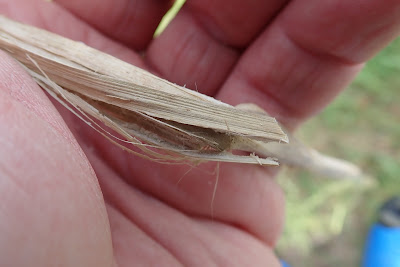
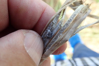
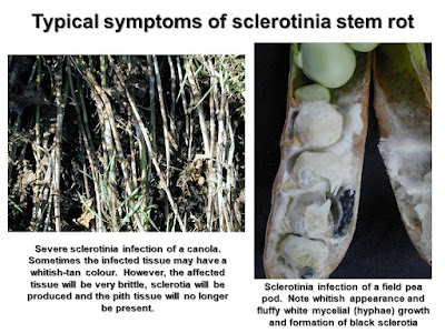
  • Infected tissue has a characteristic bleached white/whitish/tan gray appearance
  • Affected tissue when it dries is very brittle and shreds and shatters easily when you twist the stems
  • The pith portion of affected stems is typically not present
  • In heavily infected stems the pith tissue is destroyed leaving only the outer ring of stem tissue
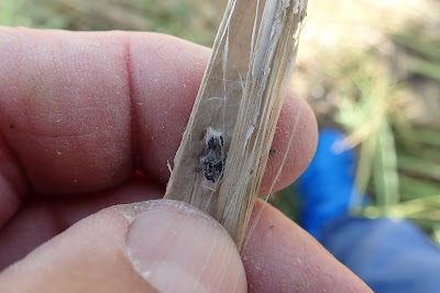
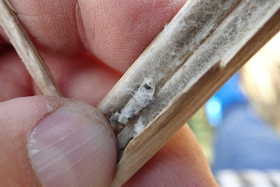
  • Sclerotia (black resting bodies) or sclerotial initials (clumps of compacted white hyphae that turn black when mature) are typically produced in or on affected plant tissues 
  • External sclerotia typically develop when you have wet conditions following infection and as the crop progresses towards senescence.


Symptoms not characteristic of sclerotinia: 
  • Infected tissue doesn’t have a bleached white/whitish grey or tan appearance 
  • Affected tissue is not brittle and doesn’t shred and shatter when you twist the stems between your hands 
  • The pith portion of stems is still intact
  • Sclerotia or sclerotial initials (dense clumps of white hyphae) are not produced in/on suspect symptoms
  • Yellowing of stems/branches may be due to other factors, such as root disease, nutrient, or weather stress, etc.


For sclerotinia one can look at the incidence of infection, and focus on the main stems and branches. Infections on the main stem and/or branches have the largest impact on yield. However, if incidence is low (<5%), the impact on yield is negligible.  


In most canola fields, plants will typically lean in a particular direction.  Use this to your advantage and work with the flow of plants.


When assessing stem rot levels, one should look at a representative sample of plants throughout the field. For example, a minimum of 4-6 sites should be looked at in a quarter section field; then at each site between 50-100 plants should be assessed.  


Half or full section fields will require more sites.  If the field is quite variable then separate assessments in specific areas may be needed, e.g. lodged versus non-lodged. 


Assessments can also be made in sprayed versus check areas to determine the usefulness and benefit of in-crop fungicide applications.  Unsprayed checks, even small areas, can be quite useful.


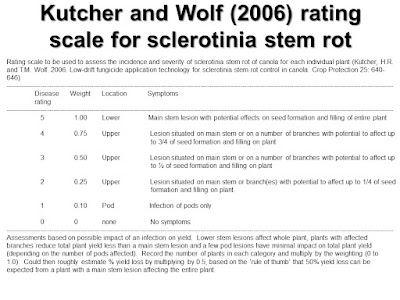
An alternative rating scale is one from Kutcher and Wolf (2006).  Although a bit more complicated, it covers infections on various parts of the plant.  Note symptoms in the upper canopy (pods, etc.) generally have less impact on yield.


Rules of thumb have been developed to assess the potential loss associated with stem rot infections.  In general the % yield loss is approximately 0.5 times the % of infected plants (main stem or main branch infections).  Thus, if you note 30% infected plants, the potential yield loss is approximately 15%.  

The PCDMN has also produced overviews for assessment of stem rot levels and identification of disease symptoms accessible with the following hyperlinks: 

• An assessment protocol for sclerotinia stem rot in canola 

• A slide deck overview of a guide to scouting and identification of sclerotinia stem rot in canola 

• Playing cards describing sclerotinia stem rot of canola symptoms

End-of-season assessment of cereal leaf spots in wheat and barley (PCDMN)

The spring and summer of 2023 was challenging for Prairie producers who experienced very dry conditions, especially under dryland production.  Although this limited disease issues, the drought stress had a very significant negative impact on stand establishment, crop growth, and yield.  However, some Prairie regions did receive moisture during the course of the summer and in these regions field crop diseases may be more of a concern.

 Although the period for scouting, risk assessment, and fungicide use is drawing to a close for cereal leaf spots in 2023,  producers and consultants may want to look at end-of season assessments. Late season crop scouting is critical for assessing the prevalence, severity and impact of these disease issues. Moreover, where unsprayed check strips or areas have been left in the field, late season assessments can be used to assess the impact and benefit of spraying in relation to leaf spot management and crop productivity.

The Prairie Crop Disease Monitoring Network (PCDMN) has developed recommended protocols for assessment of cereal leaf spots and estimates of potential yield loss.  In addition, the PCDMN has developed information on the identification of cereal leaf spot diseases.  

The main leaf spot diseases in wheat include: 

  1. Tan spot caused by the fungus Pyrenophora tritici-repentis;
  2. Speckled leaf blotch aka septoria tritici blotch caused by the fungus Zymoseptoria tritici aka Septoria tritici aka Mycosphaerella graminicola;
  3. Parastagonospora leaf and glume aka septoria nodorum leaf and glume blotch caused by the fungus Parastagonospora nodorum aka Stagonospora/Septoria nodorum;
  4. Spot blotch caused by the fungus Bipolaris sorokiniana aka Cochliobolus sativus.  It can be found in the Prairie region, but it tends to be more of a minor issue for wheat crops.  

Tan spot


Tan spot


Tan spot


Speckled leaf blotch


Speckled leaf blotch


Glume blotch.  Photo courtesy of Dr. Jeannie Gilbert


Glume blotch.  Photo courtesy of Dr. Jeannie Gilbert


Glume blotch


Glume blotch


Leaf/glume blotch


In barley, the main leaf spots include: 

  1. Scald caused by the fungus Rhynchosporium secalis aka Rhynchosporium commune.  Scald tends to be more of a cooler region disease affecting barley mainly in the cooler moister regions of Alberta;
  2. Net-form net blotch caused by the fungus Pyrenophora teres f. teres;
  3. Spot-form net blotch caused by the fungus Pyrenophora teres f. maculans;
  4. Spot blotch caused by the fungus Bipolaris sorokiniana aka Cochliobolus sativus.
Early symptoms of scald


Mature symptoms of scald


Early symptoms of net-form net blotch


Progressing symptoms of net-form net blotch

Mature symptoms of net-form net blotch


Early symptoms of spot-form net blotch


Mature symptoms of spot-form net blotch

Spot blotch

Spot blotch

One complicating factor with cereal leaf spots is potential confusion with symptoms of bacterial leaf streak in both wheat and barley.  The PCDMN also has some information on BLS that can be used to differentiate this disease from the fungal leaf spot complex in cereals.


Bacterial leaf streak of barley.  Note bacterial ooze, greasy appearance of leaf


Bacterial leaf streak of barley.  Note bacterial ooze, greasy appearance of leaf


Bacterial leaf streak of barley.  Note dried bacterial ooze (glazed doughnut appearance), greasy appearance of leaf

Bacterial leaf streak of barley.  Note dried bacterial ooze (glazed doughnut appearance), greasy appearance of leaf


Bacterial leaf streak of barley.  Note dried bacterial ooze (glazed doughnut appearance), greasy appearance of leaf


Access the Cereal Leaf Spot Assessment Protocols as a downloadable PDF file. 


Access diagrammatic images of different levels of cereal leaf spots using these  hyperlinks: 
• Standard area diagrams of wheat leaf to assess percent stripe rust
• Standard area diagrams of barley leaf to assess net-form net blotch
• Standard area diagrams of barley leaf to help assess scald

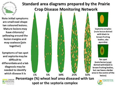
The use of standard area diagrams can help in terms of determining the level of leaf spot development.  Here are some standard area diagrams showing 1, 5, 10, 25, and 50% of the leaf area affected with various leaf spot diseases in barley and wheat.  Note that symptoms of spot-form net blotch and spot blotch in barley can be difficult to distinguish without laboratory testing of plant samples.  In addition, differentiating tan spot from the septoria complex in wheat can be challenging even for experienced plant pathologists, and thus a lab diagnosis may be needed.  However, all cereal leaf spot diseases result in destruction of leaf tissues and thus affect grain yield and filling.  The goal should be correct identification of leaf spot disease issues and then assessment of the overall level, i.e. severity. 


Use the following list of hyperlinks to gain access to PCDMN resources for cereal leaf spot identification:

Wheat:

·     Speckled leaf blotch

·     Parastagonospora/Septoria leaf and glume blotch

·    Tan spot

·     Fusarium head blight

·     Fusarium head blight seed infections

·     Bacterial leaf streak

·     Cereal rusts


Barley:

·     Net-form net blotch in barley

·     Spot-form net blotch in barley

·     Scald in barley

·     Spot blotch in barley

·     Fusarium head blight

·     Fusarium head blight seed infections

·     Cereal rusts