Introduction
Sooty moulds
Seed testing results
Fusarium head blight and fusarium damaged kernels
Wheat and barley disease information cards
Bacterial leaf streak
Introduction
With maturity
and harvest, one may assume that there are no further opportunities to obtain
plant disease information from your 2022 crop.
However, checking harvested grain for disease issues will allow you to
assess potential impacts on grade and the presence of mycotoxins. In addition, if you are planning on using
some of the harvested grain for seed then testing the grain can help to
identify potential seed health issues for the 2023 growing season.
Sooty moulds
In challenging
years when wet conditions delay harvest (e.g. 2016 and 2019), one issue that
can be of concern is the development of sooty moulds. Sooty moulds (or molds) are due to
saprophytic fungi including Alternaria
and Cladosporium species. These fungi grow on dead plant tissues when sufficient
moisture is present. Delayed harvest and
wet conditions can result in your crop going from a nice golden brown colour to
a dusty charcoal black or dark blackish olive green colour, which is due to fungal
growth on mature dead tissues. If
conditions are dry and harvesting is not delayed, sooty mould issues are
typically limited. Typical symptoms of sooty mould are shown below.
 |
| Sooty mould symptoms on ripened wheat head tissues. |
 |
| Sooty mould symptoms on ripened wheat head tissues. |
 |
| Sooty mould symptoms on ripened wheat tissues. |
 |
| Sooty mould symptoms on ripened wheat head tissues. |
Sooty
moulds can also occur on prematurely senesced plant tissues. Premature ripening within a field can be due
to abiotic (non-living) or biotic (living) factors. Abiotic factors include extreme heat stress,
frost, flooding in low areas during the growing season, etc. In contrast, biotic factors that cause
premature ripening include severe disease development especially due to root
rots (e.g. take-all root rot of wheat, clubroot of canola, Aphanomyces of field
peas). Insect damage due to wheat stem
maggot, etc. can also be responsible for premature plant ripening.
 |
| Prematurely ripened wheat plants due to take-all root rot. |
 |
| Take-all root rot symptoms in wheat showing charcoal black discolouration of lower stem bases. |
 |
Take-all root rot symptoms in wheat showing prematurely ripened tissues due to charcoal black discolouration of lower stem bases.
|
 |
| Take-all root rot symptoms in wheat showing significant root tissue destruction and charcoal black discolouration of the remaining root tissues and lower stem bases. |
 |
| Take-all root rot symptoms in wheat showing significant root tissue destruction and charcoal black discolouration of the remaining root tissues and lower stem bases. |
 |
Sooty mould development on prematurely ripened wheat heads due to take-all root rot.
|
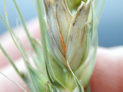
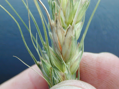
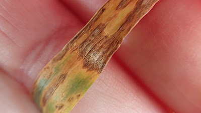
Sooty mould
development doesn't necessarily result when you have complete plant death. These pictures show sooty mould development
(blackish growth) on wheat head tissues killed due to fusarium head
blight. Note the orange/pinkish growth (sporulation
due to the rain-splashed spore stage) of the fusarium pathogen.
 |
| Early stages of sooty mould development (blackish to dark olive green fungal growth) on prematurely ripened wheat head tissue. Note the absence of fusarium head blight symptoms. |
 |
| Sooty mould development (blackish to dark olive green fungal growth) on prematurely ripened wheat spikelet tissues due to fusarium head blight. Note the presence of orange-pinkish sporulation due to the asexual rain-splashed spore stage of one or more Fusarium pathogens. |
 |
| Sooty mould development (blackish to dark olive green fungal growth) on prematurely ripened wheat spikelet tissues due to fusarium head blight. Note the presence of orange-pinkish sporulation due to the asexual rain-splashed spore stage of one or more Fusarium pathogens. |
 |
| Very early development of sooty mould development (blackish to dark olive green fungal growth) on prematurely ripened wheat spikelet tissues due to fusarium head blight. Note the presence of orange-pinkish sporulation due to the asexual rain-splashed spore stage of one or more Fusarium pathogens. |
Seed testing results
Over the
fall of 2022 and winter of 2023 you may be looking at seed testing lab results
as you prepare for the 2023 season. Of
note may be presence of Alternaria spp.
on the seed. Don't be too alarmed as it
is normal to detect Alternaria
spp. It doesn't necessarily cause seed
germination issues, although it may be indicative of weathering due to a
delayed harvest and potential development of sooty moulds. If you find levels of Alternaria spp. that range from 10-40%, but the germination level
is good (e.g. >95%), the seed should be fine for planting in 2023. However, adding a good quality seed treatment
with effective application technology can provide peace of mind and mitigate
any potential issues affecting stand establishment. Make sure to talk with your seed health
professionals on what they are finding in terms of fungal load, germination,
and vigour.
Fusarium head blight and fusarium damaged kernels
One other
issue that farmers may be seeing is downgrading in wheat and durum due to the
presence of fusarium damaged kernels in
areas where Fusarium graminearum is
well-established and you have had increased moisture during head emergence and
anthesis. This downgrading is most likely due to Fusarium graminearum and the presence of fusarium damaged kernels
(FDK), while there may be underlying mycotoxin issues. Keep in mind that other Fusarium species may be present and more commonly found especially
under drier conditions versus Fusarium
graminearum.
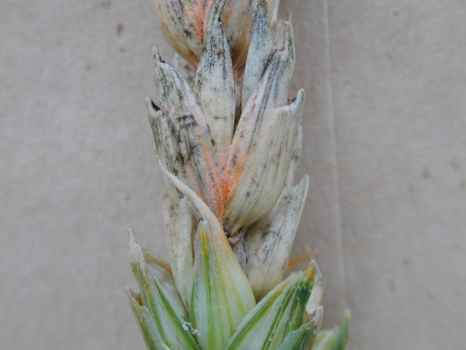
 |
| Fusarium head blight symptoms in wheat. Note the presence of orange-pinkish sporulation at the base of the spikelet due to the asexual rain-splashed spore stage of one or more Fusarium pathogens. |
 |
| Fusarium head blight symptoms in wheat. Note the presence of orange-pinkish sporulation at the base of the spikelet due to the asexual rain-splashed spore stage of one or more Fusarium pathogens. |
 |
Fusarium head blight symptoms in wheat. Note the presence of orange-pinkish sporulation in the crevices of the floret tissues of the spikelet due to the asexual rain-splashed spore stage of one or more Fusarium pathogens.
|
 |
| Fusarium head blight symptoms in wheat. Note the presence of orange-pinkish sporulation in the crevices of the floret tissues of the spikelet due to the asexual rain-splashed spore stage of one or more Fusarium pathogens. |
 |
| Fusarium head blight symptoms in wheat. Note the presence of orange-pinkish sporulation in the crevices of the floret tissues of the spikelet due to the asexual rain-splashed spore stage of one or more Fusarium pathogens. Also present is the blackish-dark olive green growth of the sooty mould fungi on the prematurely ripened tissue. |
 |
| Fusarium head blight symptoms in wheat. Note the presence of orange-pinkish sporulation in the crevices of the floret tissues of the spikelet due to the asexual rain-splashed spore stage of one or more Fusarium pathogens. |
 |
Fusarium head blight symptoms in wheat. Note the presence of orange-pinkish sporulation in the crevices of the floret tissues of the spikelet due to the asexual rain-splashed spore stage of one or more Fusarium pathogens.
|
 |
| Fusarium head blight symptoms in wheat. If conditions turn dry following infection there may not be much if any orange-pinkish sporulation in the crevices of the prematurely ripened floret tissues. However, one diagnostic feature of FHB is the browning of the rachis and peduncle tissue. |
 |
| Fusarium head blight symptoms in wheat. If conditions turn dry following infection there may not be much if any orange-pinkish sporulation in the crevices of the prematurely ripened floret tissues. However, one diagnostic feature of FHB is the browning of the rachis and peduncle tissue. |
 |
| Fusarium head blight symptoms in wheat. If conditions turn dry following infection there may not be much if any orange-pinkish sporulation in the crevices of the prematurely ripened floret tissues. However, one diagnostic feature of FHB is the browning of the rachis and peduncle tissue. |
 |
| Fusarium head blight symptoms in wheat. If conditions turn dry following infection there may not be much if any orange-pinkish sporulation in the crevices of the prematurely ripened floret tissues. However, one diagnostic feature of FHB is the browning of the rachis and peduncle tissue. |
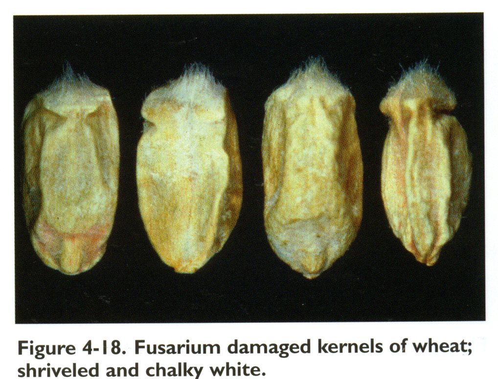
 |
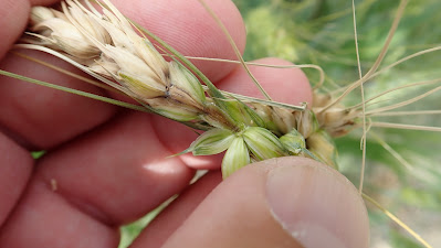
| Healthy and typical chalky white shriveled kernel symptoms, i.e. fusarium damaged kernels. |
 |
| Healthy and typical chalky white shriveled kernel symptoms, i.e. fusarium damaged kernels. |
Things
become more challenging in areas where Fusarium
graminearum is not present or present at low levels. Here the down grading you may be seeing in
your wheat/durum can be due to other issues and not necessarily Fusarium graminearum. As a consequence
mycotoxins such as deoxynivalenol aka DON may not be a concern. If you haven’t had issues with Fusarium graminearum on your farm in the
past, the presence of FDK may be due to other Fusarium spp. (e.g. F.
avenaceum) or in fact other non-Fusarium pathogens. The main non-Fusarium pathogen that can
produce FDK-like symptoms is the leaf/glume blotch pathogen, Parastagnospora nodurum aka Septoria nodorum. The glume blotch pathogen produces brownish
lesions on the glumes, but can also affect the seed. The leaf and glume blotch pathogen, Parastagonospora nodorum, aka Stagonospora nodorum/Septoria nodorum can produce FDK
symptoms that mimic those caused by Fusarium
graminearum. Figure 4-3 below is
courtesy of the Canadian Grain Commission.
 |
| Symptoms of glume blotch pathogen infection of wheat. Photo courtesy of Randy Clear (retired), Canadian Grain Commission. |
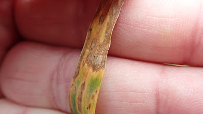
You can
note symptoms of glume blotch in your fields prior to harvest. Typical symptoms include brownish or
purple-brown lesions on glumes. Also if
you look closely enough you may be able to see small brownish bumps or pycnidia
covering the lesions. Pycnidia are asexual fruiting structures that produce the rain-splashed spore stage of the glume blotch pathogen.
 |
| Typical brownish-purply brown symptoms of glume blotch of wheat. Photo courtesy of Dr. Jeannie Gilbert (retired), Cereal Research Centre, AAFC Winnipeg. |
 |
| Typical brownish-purply brown symptoms of glume blotch of wheat. Note the presence of the asexual fruiting bodies aka pycnidia that produce the rain-splashed spore stage of the glume blotch pathogen. Photo courtesy of Dr. Jeannie Gilbert (retired), Cereal Research Centre, AAFC Winnipeg. |
 |
| Typical brownish-purply brown symptoms of glume blotch of wheat. Note the presence of the asexual fruiting bodies aka pycnidia (brownish bumps) that produce the rain-splashed spore stage of the glume blotch pathogen. |
 |
| Typical brownish-purply brown symptoms of glume blotch of wheat. Note the presence of the asexual fruiting bodies aka pycnidia (brownish bumps) that produce the rain-splashed spore stage of the glume blotch pathogen. |
 |
| Typical tan-brown lesions of the leaf blotch phase of glume blotch of wheat. Note the presence of the asexual fruiting bodies aka pycnidia (brownish bumps) that dot the lesions. These pycnidia produce the rain-splashed spore stage of the glume blotch pathogen. |
 |
| Typical tan-brown lesions of the leaf blotch phase of glume blotch of wheat. Note the presence of the asexual fruiting bodies aka pycnidia (brownish bumps) that dot the lesions. These pycnidia produce the rain-splashed spore stage of the glume blotch pathogen. |
The
Canadian Grain Commission has a great resource to illustrate typical FDK
symptoms and those caused by other fungi.
See: https://grainscanada.gc.ca/en/grain-quality/grain-grading/grading-factors/identifying-fusarium.html.
These
pictures illustrate typical grain symptoms due to Fusarium graminearum, i.e. the chalky white shriveled kernels
versus intact healthy kernels.
 |
| Typical grain symptoms due to Fusarium graminearum, i.e. the chalky white shriveled kernels versus intact healthy kernels. |
 |
| Typical grain symptoms due to Fusarium graminearum, i.e. the chalky white shriveled kernels versus intact healthy kernels. |
 |
| Typical grain symptoms due to Fusarium graminearum, i.e. the chalky white shriveled kernels versus intact healthy kernels. |
 |
| Typical grain symptoms due to Fusarium graminearum, i.e. the chalky white shriveled kernels versus intact healthy kernels. |
 |
| Typical grain symptoms due to Fusarium graminearum, i.e. the chalky white shriveled kernels versus intact healthy kernels. Note Fig. 4.18 is courtesy of Randy Clear (retired), Canadian Grain Commission. |
 |
| Typical grain symptoms due to Fusarium graminearum, i.e. the chalky white shriveled kernels versus intact healthy kernels. |
This picture is
of durum with kernels exhibiting symptoms of FDKs due to Fusarium graminearum.
However, there are also other kernels with a reddish tinge. These reddish/pinkish symptoms are typical of
the tan spot pathogen that can affect leaves, heads and grain, and are called
red smudge, which is a grading factor especially in durum wheat.
 |
| Healthy, fusarium damaged kernels (chalky white and shriveled) and reddish discoloured (red smudge due to the tans spot pathogen) durum wheat kernels. The reddish/pinkish symptoms are typical of the tan spot pathogen that can affect leaves, heads and grain, and are called red smudge, which is a grading factor especially in durum wheat. |
| Healthy, fusarium damaged kernels (chalky white and shriveled) and reddish discoloured (red smudge due to the tans spot pathogen) durum wheat kernels. The reddish/pinkish symptoms are typical of the tan spot pathogen that can affect leaves, heads and grain, and are called red smudge, which is a grading factor especially in durum wheat. |
| Healthy (middle) and and reddish discoloured (red smudge due to the tans spot pathogen) durum wheat kernels. The reddish/pinkish symptoms are typical of the tan spot pathogen that can affect leaves, heads and grain, and are called red smudge, which is a grading factor especially in durum wheat. |
 |
Typical tan-coloured lesions of the tan spot fungus of wheat and durum.
|
If you are
in an area with no to a limited history of Fusarium
graminearum, you may still have issues with downgrading due to FDK. In this case make sure to send your grain to
a seed testing laboratory to have a fungal screen done and also test for the
mycotoxin deoxynivalenol (DON). You may
find that the FDKs in your wheat are actually not due to Fusarium graminearum, but other Fusarium
spp. where DON is not an issue OR where they are due to other fungal
pathogens such as the glume blotch fungus.
Fungal screening and DON tests may indicate no Fusarium graminearum or DON, and thus you may be able to have your
grade reassessed or at least open up additional market/end-use options,
including using the grain for hog feed.
Wheat and barley disease information cards
The PCDMN
has developed disease info cards for fusarium head blight of wheat and barley
caused Fusarium graminearum. If you observed symptoms of FHB in your wheat
and barley in late July and early August make sure to closely check the
harvested grain for FHB symptoms. Again
having suspect grain tested by a seed testing lab can provide information that
may help with grading and mycotoxin issues, while providing additional marketing
options. The PCDMN has also prepared a disease
info card that outlines typical fusarium damaged kernel (FDK) symptoms as well
as symptoms due to other issues that may be confused with fusarium head blight
damage in harvested grain.
See:
PCDMN Disease info cards for wheat:
·
Speckled leaf blotch
·
Parastagonospora/Septoria leaf and glume blotch
·
Tan spot
·
Fusarium head blight
·
Fusarium head blight seed infections
·
Bacterial leaf streak
·
Cereal rusts
Bacterial leaf streak
One
additional concern in cereals in relation to seed and crop health is bacterial
leaf streak, which is becoming an increasing concern for Prairie cereal
producers. The PCDMN has developed a
disease info card on bacterial leaf streak as well as posting some additional
information earlier this summer.
See:
https://twitter.com/pcdmn/status/1555320801670438913
https://twitter.com/pcdmn/status/1522349120119259137
https://twitter.com/pcdmn/status/1546478451015778304
https://drive.google.com/file/d/1Z9YmfHLaYQ8qqejofUpDafy7mq8qmfQp/view?usp=sharing


